


Eye and Ear Infections: Engaging with NotebookLM
Join NotebookLM—study smarter, cite every fact!

Prologue – Tyler & Maya Board the Flight Deck
Tyler still remembers the dopamine hit of Module’s Microbiology Gold Cup micro-quests that turned his thumb into a flash-card machine-gun. Maya could chant Streptococcus Lancefield groups in her sleep, too, but both sensed the same gap: real patients don’t arrive as disembodied trivia.
So when their shared screen lights up with a new mission—
“Eye & Ear Infections: Engaging with NotebookLM”—they lean in.
Welcome to the next level of micro-learning. Instead of juggling stray lecture slides and half-forgotten pharm tables, Tyler drags today’s otitis-media deck, Maya adds yesterday’s amoxicillin-clavulanate cheat-sheet, and NotebookLM weaves a single, patient-ready storyline—from a toddler’s bulging eardrum to the red-eye they’ll meet on rounds tomorrow.
Inside the notebook they can:
Bundle & compare sources—shared mechanisms pop, sneaky contradictions surface in seconds.
Viva-voce on demand—“Quiz me like an OSCE examiner,” answers graded with cited lines.
Podcast recap—hit Audio Discussion and a five-minute talk-show summary drops into Tyler’s treadmill queue.
Map the gaps—NotebookLM flags thin subtopics; Maya turns them into the next Anki deck.
Flashcards, done—one prompt converts the pharm table into more flash cards for spaced repetition.
Eight-second swipes still feed the dopamine loop—only now each swipe propels a clinical narrative.
Grab the notebook, sync your sources, and turn eight-second swipes into bedside fluency. Game on!
Click here - Here is the NotebookLM

Dear Future Physician,
You’ve just opened a notebook that thinks with you, not for you. NotebookLM isn’t another “upload-and-summarize” gadget—it’s a clinical reasoning partner designed to help you connect, question, and master the mountain of documents you juggle in medical school.
🚀 Dive in—Happy exploring, and may your curiosity drive better patient care!
HEENT (Eye and Ear) Infections:
These sources provide a comprehensive overview of common ear and eye infections, focusing on etiology, clinical presentation, and management strategies. They present detailed lists of prevalent pathogens associated with various conditions like otitis media, conjunctivitis, and keratitis, often ranked by frequency. The texts also explain the pathogenesis of these infections, outlining the mechanisms by which pathogens cause disease and how host factors influence susceptibility. Furthermore, they offer high-yield clinical pearls and mnemonics, emphasizing critical diagnostic clues and first-line treatments relevant for medical students.
Audio Overview:
Want more details, read on, better still, go straight to NotebookLM and ask questions:
Eye Infections
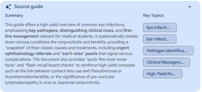
EYE INFECTIONS
A. Conjunctivitis
Adherence: Bacterial adhesins (S. aureus, H. influenzae) or viral fiber proteins (adenovirus) bind conjunctival glycocalyx.
Barrier breach: Toxins or lytic viral replication disrupt tight junctions, increasing vascular permeability.
Cytokine cascade (IL-1β, TNF-α) recruits neutrophils → purulent discharge (bacterial) or lymphoid follicles with pre-auricular node (viral).
Allergic form: IgE-mediated mast-cell degranulation releases histamine—edema, itching, “cobblestoning.”
B. Keratitis (Corneal Ulcer)
Epithelium disruption (contact-lens hypoxia, trauma) exposes stromal collagen.
Bacterial toxins: Pseudomonas elastase, exotoxin A, and biofilm degrade stroma at lightning speed.
Fungal hyphae (Fusarium, Aspergillus) penetrate vertically, producing feathery infiltrates and satellite lesions.
Acanthamoeba trophozoites encyst in stromal lamellae; radial neuritis explains exquisite pain.
Host response: Neutrophil metalloproteinases and complement amplify melting → descemetocele or perforation.
C. Endophthalmitis
Entry routes: intra-operative contamination (CoNS), penetrating trauma (Bacillus cereus), or hematogenous seeding (Candida).
Vitreous growth chamber: nutrient-rich, avascular gel permits exponential microbial replication.
Inflammatory surge: IL-6, IL-8, and vitreous collagen breakdown form hypopyon; blood–retina barrier disruption causes painless vision drop then severe ache.
Toxin-mediated necrosis: B. cereus hemolysins and cereolysin accelerate retinal destruction within hours.
D. Lacrimal-Apparatus Infections
Stasis in obstructed ducts (nasolacrimal stenosis) predisposes to S. aureus or Strep dacryocystitis; pressure drives bacteria retrograde into sac mucosa.
Actinomyces canaliculitis forms sulfur-granule concretions that perpetuate low-grade inflammation until mechanically removed.
Shared principles: loss of anatomic barriers (tear film, corneal epithelium), microbial virulence factors (adhesins, exotoxins, biofilm), and the eye’s confined spaces together dictate the speed and severity of ocular infection and the narrow window for vision-saving intervention.
EAR INFECTIONS
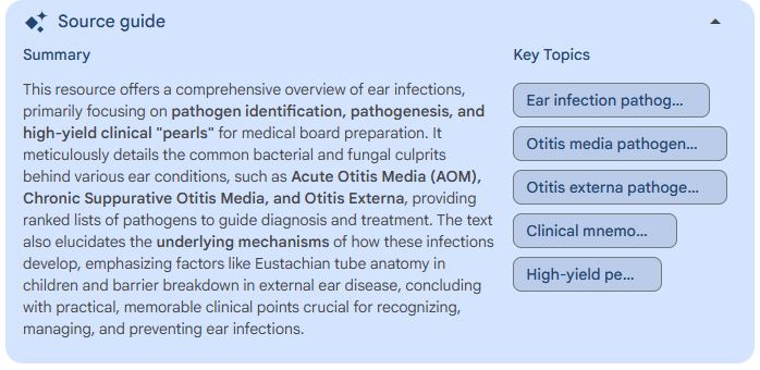
Middle-Ear Disease (Acute & Chronic Otitis Media)
Mucosal trigger: Most episodes start with a viral URI; edema of the nasopharynx and Eustachian tube (ET) blocks aeration and generates negative middle-ear pressure that wicks colonizing bacteria inward.
Pediatric anatomy & immunity: Children’s ETs are shorter, more horizontal, and lack mature cartilaginous tone, while waning maternal IgG and limited endogenous antibodies weaken mucosal defense.
Fluid → microbe niche: Sterile transudate trapped behind the tympanic membrane becomes a nutrient medium where nontypeable H. influenzae, S. pneumoniae, and M. catarrhalis proliferate; pneumococcal capsule triggers robust neutrophilic inflammation, explaining higher fevers and mastoid risk.
Mucosal spread variant: H. influenzae readily colonizes adjacent conjunctiva, producing the otitis–conjunctivitis syndrome seen in about 10 % of cases.
Chronic suppuration: Repeated pressure or acute perforation converts AOM to a “wet ear.” The aerated cavity favors P. aeruginosa and S. aureus with persistent discharge, keratin debris, and osteitic change.
External-Ear Disease (Otitis Externa Spectrum)
Barrier breakdown: Canal skin loses its acidic, hydrophobic shield when repeatedly wet (swimming), traumatized by Q-tips, or inflamed by eczema/dermatitis, enabling bacterial adherence.
Local inflammation loop: P. aeruginosa and S. aureus secrete proteases and exotoxins that damage keratin, raise pH, and swell the canal; edema plus exudate intensifies pain on tragus or pinna traction.
Fungal takeover: After topical antibiotics or in humid climates, Aspergillus or Candida exploit the disrupted flora to cause pruritic, black-or-white-spored otomycosis.
Malignant progression: In diabetes or immunosuppression, P. aeruginosa invades through fissures of Santorini into the skull base, producing osteomyelitis and cranial-nerve VI–XII palsies; high-resolution CT confirms bone erosion.
Chronic external otitis: Persistent moisture, allergic contact dermatitis, or psoriasis sustains low-grade inflammation, granulation tissue, and eventual canal stenosis if not corrected.
These mechanistic steps connect anatomy, host defenses, and pathogen virulence, equipping learners to predict complications and choose targeted interventions.
Epilogue – Credits Roll, Controllers Ready
Tyler’s phone buzzes one last time before the weekend.Achievement Unlocked: “Watery “gritty” red eye → Clear Thinking.”
Maya cracks her knuckles.
Faculty queue coaching clips instead of crafting quizzes.
Game (still) on.
My Dear Students Physicians, this is my first attempt with NotebookLM. Please share your experience with me and how I can improve the experience. Email me or write in the comments box, below.
Disclaimer
This material is provided exclusively for undergraduate medical education. It does not constitute medical advice, diagnosis, or management guidance for eye or ear infections. The author and contributors accept no responsibility for clinical decisions or patient outcomes. Always consult up-to-date evidence-based references and licensed healthcare professionals before applying any information in practice.